

|
Overview |
||
|
Product Name |
Iba1/AIF1 antibody |
|
|
Product Number |
||
| Gene Description |
Allograft inflammatory factor 1 |
|
| Clonality |
Polyclonal |
|
| Host |
Rabbit |
|
| Species Reactivity |
Human, mouse, rat |
|
| Recommended Applications Dilutions |
Immunofluorescence 1:100 – 1:300 Immunohistochemistry (Paraffin) 1:100 – 1:300 Immunohistochemistry (Frozen) 1:100 – 1:300 |
|
| Storage Buffer |
100mM Tris Glycine, 1% BSA, 20% Glycerol (pH7). 0.025% ProClin 300 was added as a preservative |
|
| Concentration |
0.7 mg/ml |
|
| Purity |
Affinity column purified |
|
| Storage |
Store at +4°C for short term storage. Long time storage is recommended at -20°C |
|
|
Notes |
Gently mix before use. Optimal concentrations and conditions for each application should be determined by the user. |
|
|
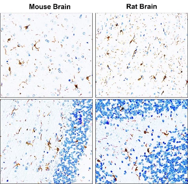
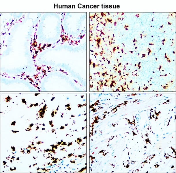
IR13-36 anti-Iba1/AIF1 antibody IHC image |
IR13-36 anti-Iba1/AIF1 antibody IHC image |
|
 |
 |
|
|
Immunohistochemical analysis of paraffin embedded Mouse / Rat brain tissue labeling Iba1 with IR13-36 at 1/200. Epitope Retrieval methods: Citrate Buffer, pH 6.0 Data from: Li-Tzung Biotechnology. Co |
Immunohistochemical analysis of paraffin embedded Human cancer tissue labeling Iba1 with IR13-36 at 1/200. Epitope Retrieval methods: Citrate Buffer, pH 6.0 |
|
|
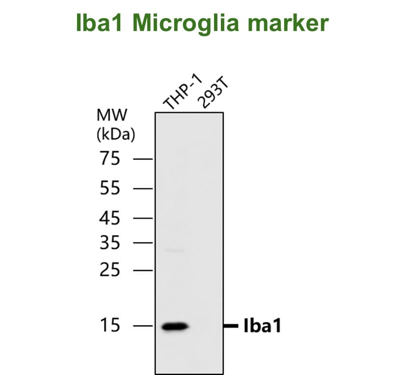
IR13-36 anti-Iba1/AIF1 antibody WB image |
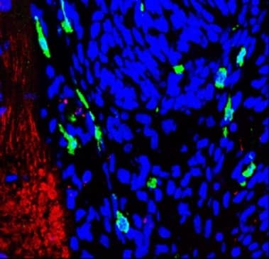
Customer feedback Image IR13-36 anti-Iba1/AIF1 antibody IHC-Fr image |
 |
 |
|
Iba1/AIF1 antibody - at 1/500 dilution Lysates at 45μg per lane, This blot was produced using a 15% SDS-PAGE. PVDF membrane was then blocked with 3%BSA for an hour before being incubated with IR13-36 overnight at 4°C. |
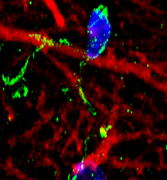
Frozen sectioned for immunohistochemical analysis.
Sample: Frozen sectioned Rat brain. Green: Iba1 antibody (IR13-36) Red: TUJ1 antibody Blue: DAPI |
|
Customer feedback Image IR13-36 anti-Iba1/AIF1 antibody IHC-Fr image |
Customer feedback Image IR13-36 anti-Iba1/AIF1 antibody IHC-Fr image |
 |
 |
|
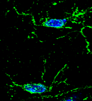
Immunohistochemical of frozen sections. Sample : mouse cerebellum. Green : Iba1 antibody (IR13-36) : 1-200 Anti-rabbit 488 : 1-500. DAPI (blue) was used as the nuclear counter stain. |
Immunohistochemical of frozen sections. Sample : mouse hippocampus. Green : Iba1 antibody (IR13-36) : 1-200 Anti-rabbit 488 : 1-500. DAPI (blue) was used as the nuclear counter stain. |